X-ray tomography
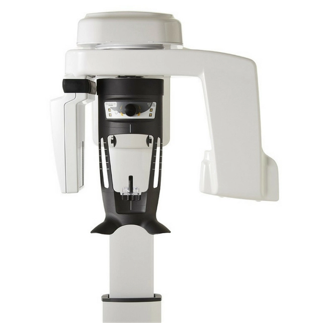
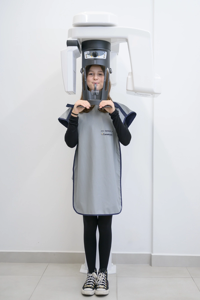
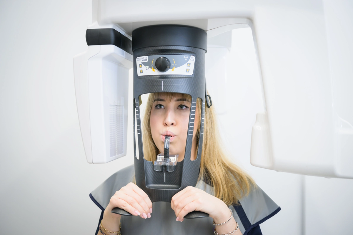
We invite you to the Implant-Art office, where diagnosis and treatment are supported by the latest equipment for taking panoramic x-rays and computer tomography of the jaws.
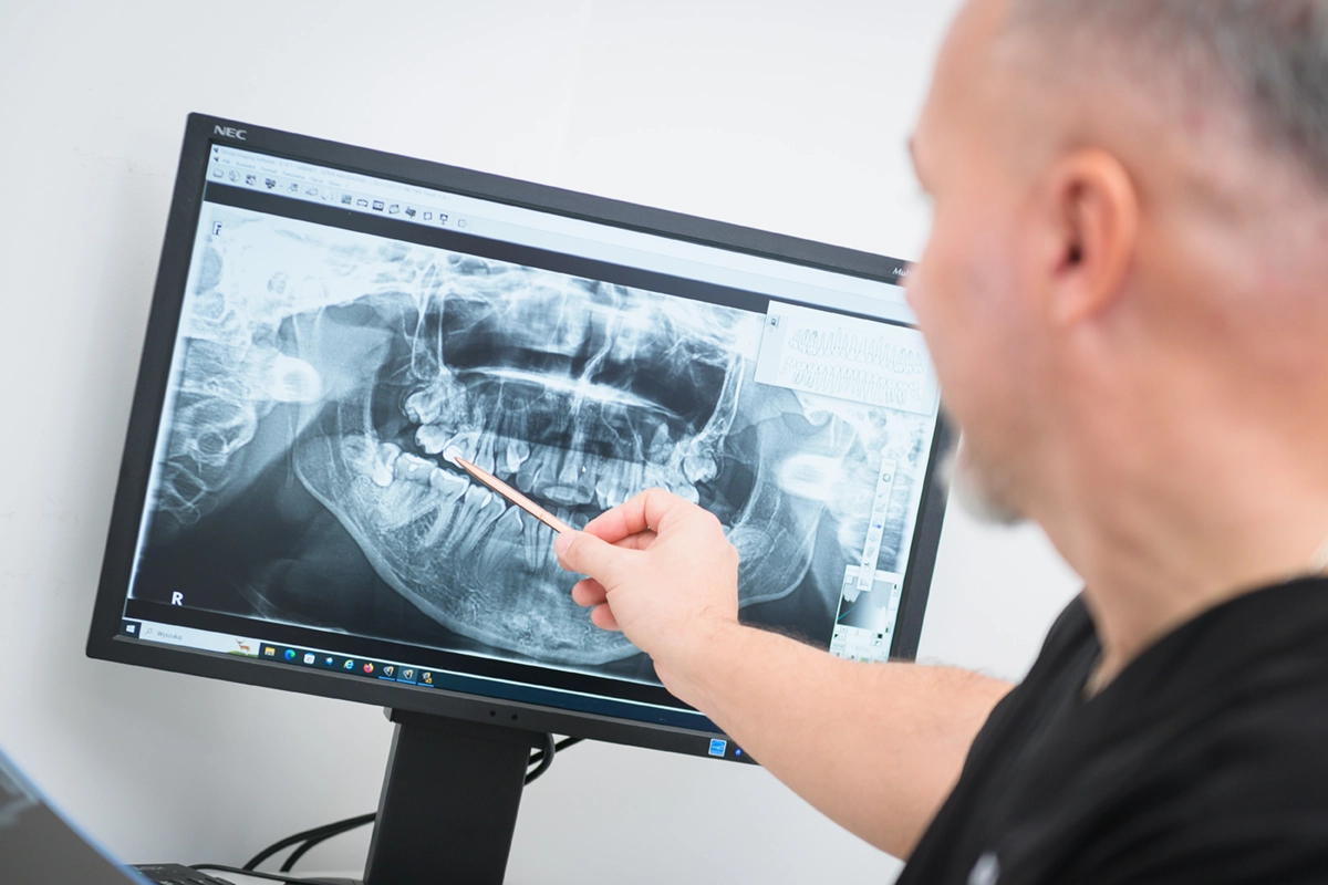
Thanks to the panoramic radiograph, we obtain a complete picture of the teeth, including malocclusions, impacted teeth, or pathological changes that cannot be assessed during a clinical examination.
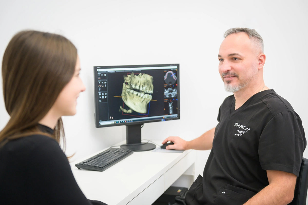
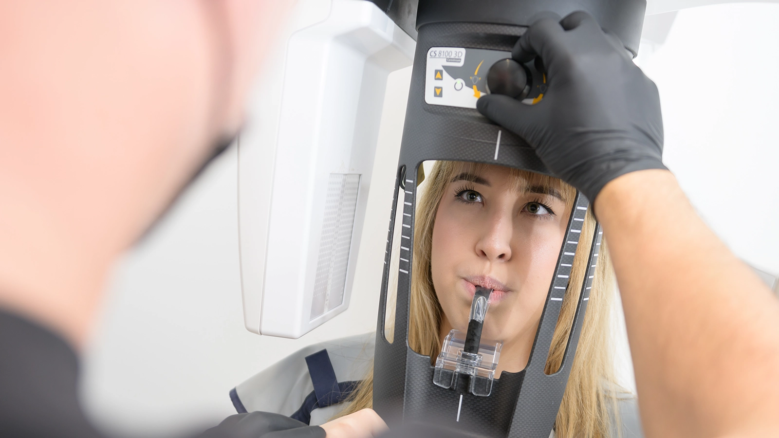
In the case of computed tomography of the jaws, the imaging field may cover both one tooth and the entire maxilla and/or mandible.
The computed tomography of the jaws performed in our office, thanks to its three-dimensional image, is used in many fields of dentistry, including: in implantology, surgery and endodontics.
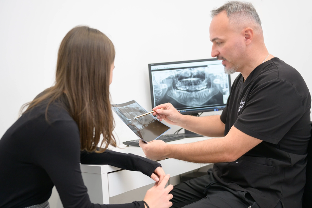
In implantology, it allows us to assess the width and height of the bones and the location of anatomical structures in the cross-section or plane of interest. Thanks to this, it is possible to precisely determine the position of the implant and precisely plan the procedure. This test is also used in specialized computer programs that simulate a dental implant.
In surgery, computed tomography enables accurate localization of impacted teeth (eights) and anatomical structures. Thanks to this, for example, surgical removal of wisdom teeth is even more precise and the procedure time is shorter.
In the case of endodontics, tomography helps find additional canals and locate broken instruments.
In orthodontics – it enables precise planning of treatment for malocclusions.
In our office, we also use computed tomography to diagnose the maxillary sinuses and temporomandibular joints.
Due to the fact that we use the most modern equipment for the diagnostics described above, the radiation dose is reduced to a minimum and the advantages are indisputable. Therefore, we invite you to professional diagnosis and treatment.